

Skip to content
View in the app

A better way to browse. Learn more.

Pediatric Surgery Club

A full-screen app on your home screen with push notifications, badges and more.

To install this app on iOS and iPadOS
  1. Tap the Share icon in Safari
  2. Scroll the menu and tap Add to Home Screen.
  3. Tap Add in the top-right corner.
To install this app on Android
  1. Tap the 3-dot menu (⋮) in the top-right corner of the browser.
  2. Tap Add to Home screen or Install app.
  3. Confirm by tapping Install.
Get your Pro Membership!

admin

Administrators
  • Joined

  • Last visited

Everything posted by admin

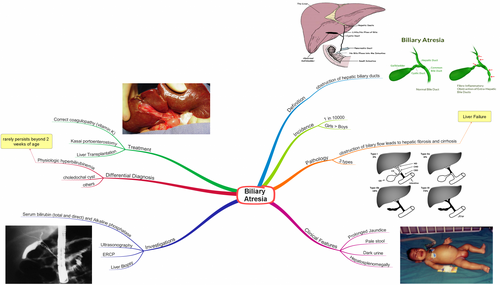
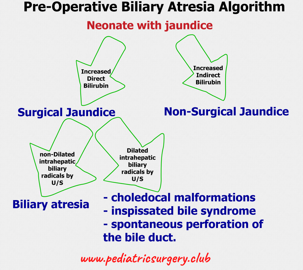
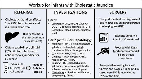
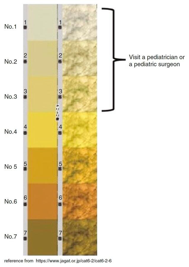
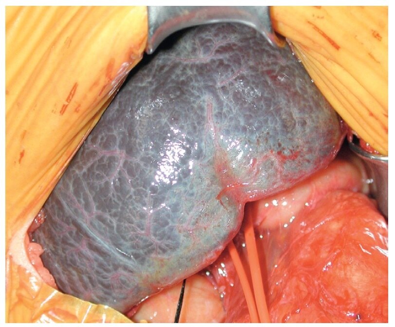
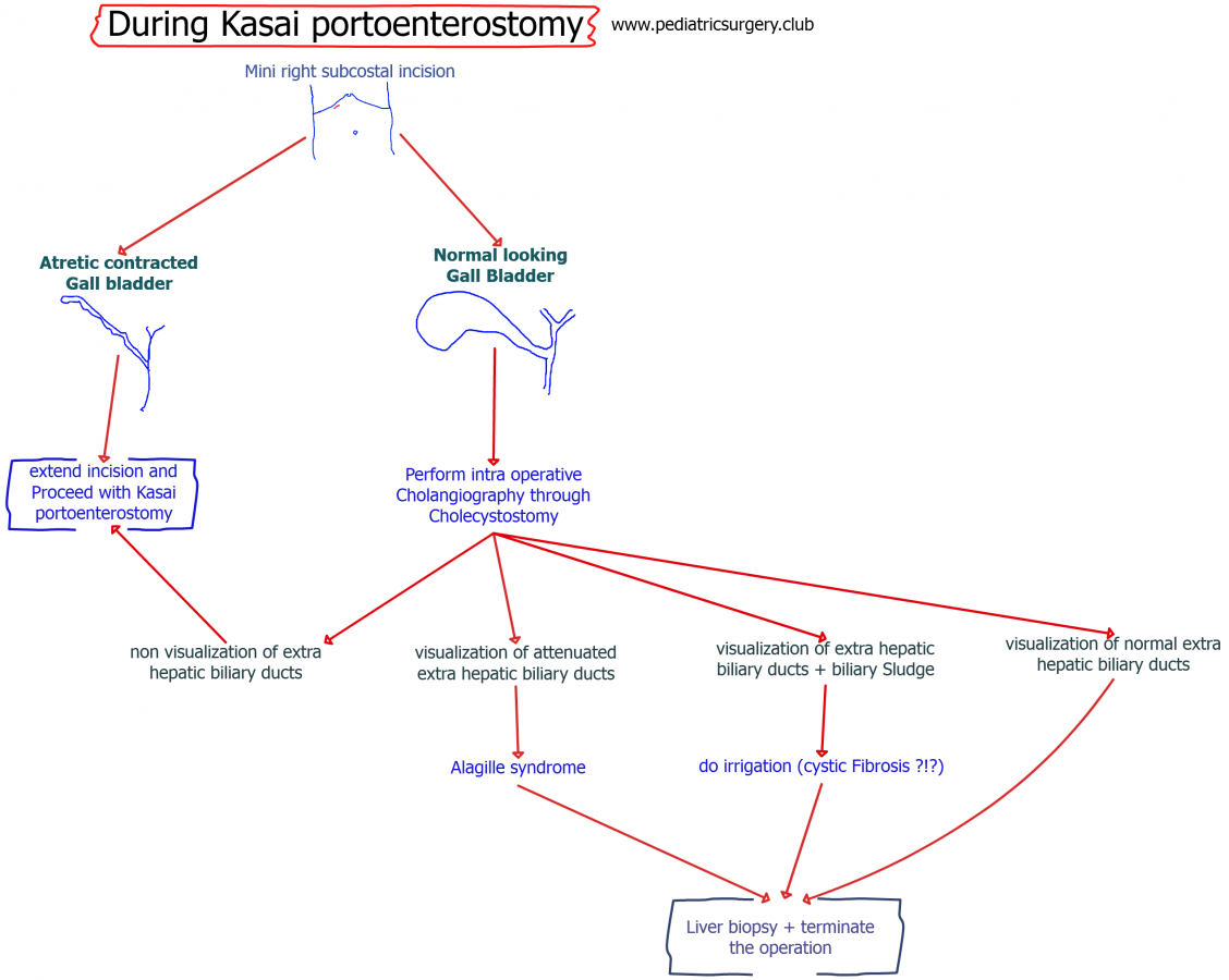
  1. Quote from "The SAGES Manual of Pediatric Minimally Invasive Surgery" by Danielle S. Walsh, Todd A. Ponsky, Nicholas E. Bruns - "The onset of jaundice may occur at birth or up to 6 weeks thereafter and is typically progressive and eventually accompanied by acholic stools and dark urine." Quote from "The SAGES Manual of Pediatric Minimally Invasive Surgery" by Danielle S. Walsh, Todd A. Ponsky, Nicholas E. Bruns - "Hepatic scintigraphy (HIDA scan) demonstrating relatively good hepatic uptake with the absence of excretion of technetium-labeled compounds from the liver into the duodenum is diagnostic of biliary obstruction, but may be less reliable in cases of severe jaundice. Prior to HIDA scan, patients are routinely pretreated with phenobarbital (5 mg/kg/day) for 5 days to increase biliary secretion by stimulating hepatic enzymes to minimize studies." Quote from "The SAGES Manual of Pediatric Minimally Invasive Surgery" by Danielle S. Walsh, Todd A. Ponsky, Nicholas E. Bruns - "biopsy of the liver is the most accurate diagnostic test, but also the most invasive, short of surgical exploration [23, 24]. Pathology characteristically reveals inflammation with ductular proliferation, with or without bile stasis, duct plugging, and giant cells. Indeterminate or false-negative results may reflect specimens acquired too early in the disease process to reflect the characteristic pathologic changes of duct proliferation."
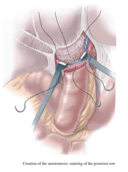
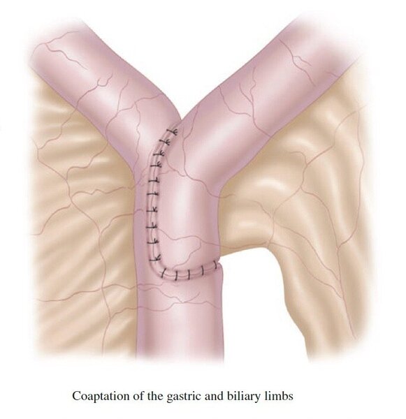
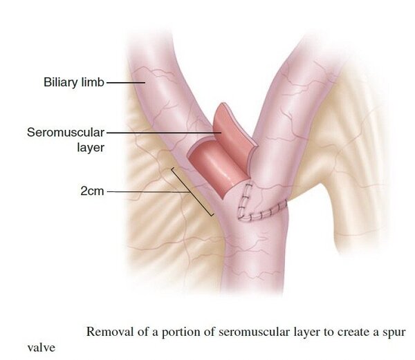
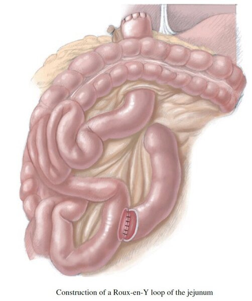
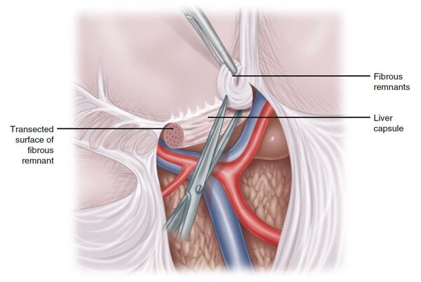
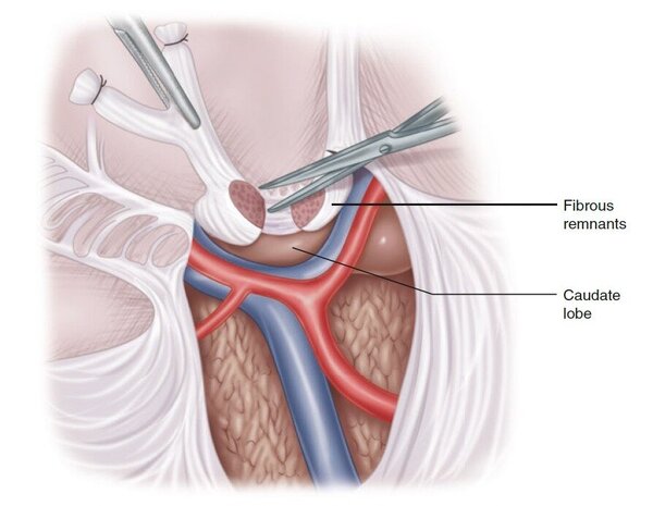
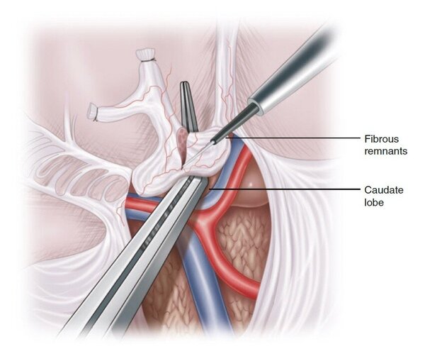
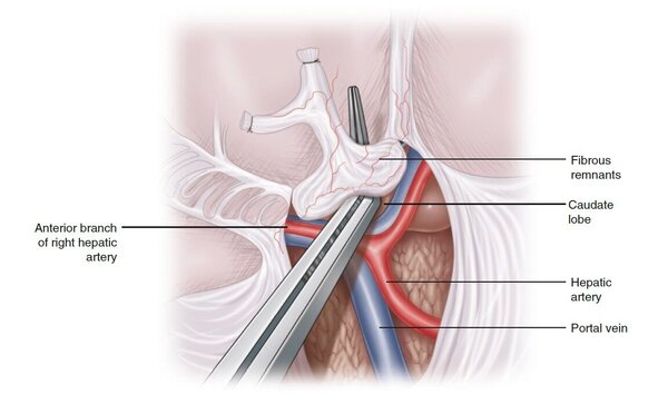
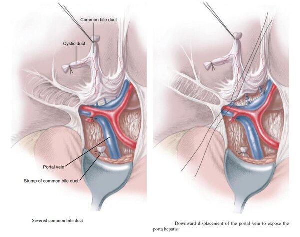
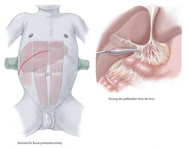
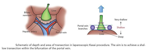
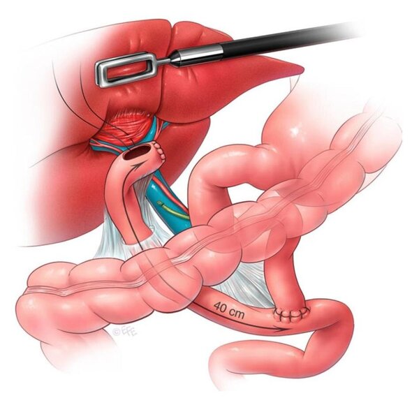
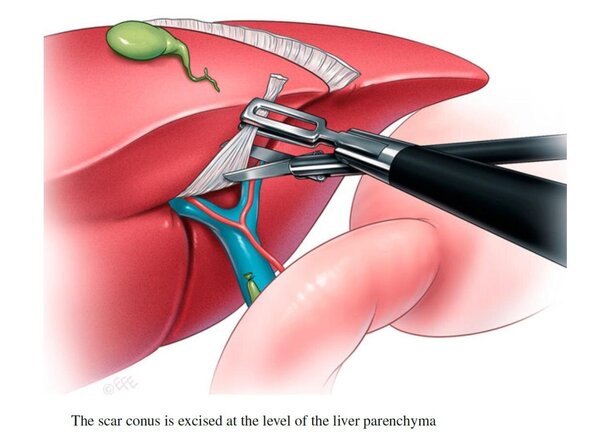
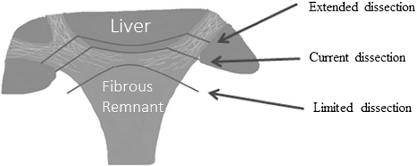
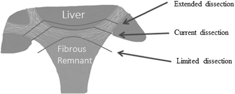
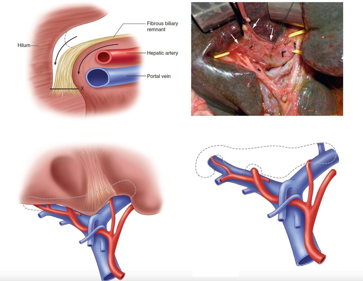
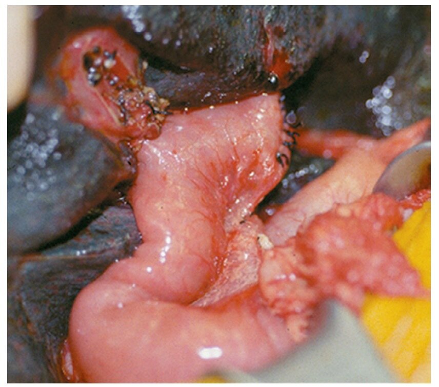
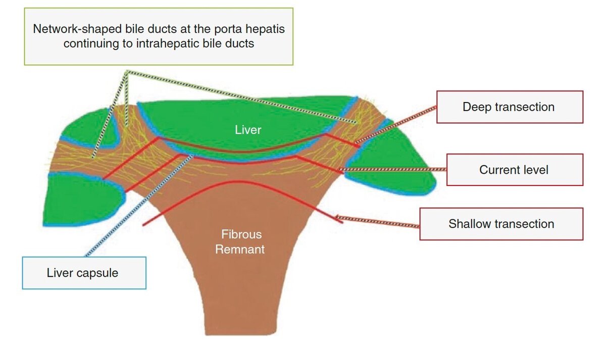
  2. Quote from "The SAGES Manual of Pediatric Minimally Invasive Surgery" by Danielle S. Walsh, Todd A. Ponsky, Nicholas E. Bruns - "The key of the procedure is dissection of the cone-shaped fibrous remnant at the level of the liver surface, just anterior to the portal vein bifurcation. For best long-term results, the bilious scar tissue must be removed directly on the liver surface without injuring the actual liver. The dissection must be carried as much lateral as possible with typical limitations of right and left portal venous and hepatic artery branches as well as below the portal vein bifurcation. The completely exposed liver surface at the porta hepatis is then covered by the Roux limb as a quasi-“sewer” to catch dripping bile from the area."
  3. Quote from "The SAGES Manual of Pediatric Minimally Invasive Surgery" by Danielle S. Walsh, Todd A. Ponsky, Nicholas E. Bruns - "Trendelenburg positioning may also augment contrast delivery to the intrahepatic bile ducts if initial images are equivocal, and administration of intravenous glucagon may improve drainage of contrast into the duodenum if such flow is not initially evident"
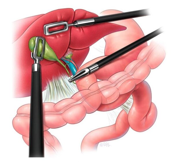
  4. Quote from "ESPES Manual of Pediatric Minimally Invasive Surgery" by Ciro Esposito, François Becmeur, Henri Steyaert, Philipp Szavay - "The first ever cholecystectomy on record was performed on July 15, 1882, by Carl Johann August Langenbuch (1846–1901) at Lazarus Krankenhaus in Berlin [1]. Few years earlier, on July 15, 1867, John Stough Bobbs of Indianapolis had performed a cholecystostomy on a 30-year-old woman with ovarian cancer [2]. It was over 100 years later, in Germany again, that Erich Mühe of Boblingen performed the first laparoscopic (Fig. 37.1) cholecystectomy [3]. This was a turning point for minimal access approach in general surgery. Another major advance, which helped the surgeons, was the development of a laparoscopic clip applicator with multiple clips. Dr. Mühe had to overcome a lot of hurdles before his technique was being recognised and adopted by other surgeons. He submitted his article to The American Journal of Surgery in 1990 but was rejected. His article never got published in English literature. The procedure was performed on September 12, 1985, with the help of a side-viewing endoscope and an instrumentation channel through the umbilicus. Eventually, his work was rewarded by him receiving the German Surgical Society Anniversary Award with praises like “One of the greatest achievements of German medicine in recent history.”"

Account

Navigation

Search

Search

Configure browser push notifications

Chrome (Android)
  1. Tap the lock icon next to the address bar.
  2. Tap Permissions → Notifications.
  3. Adjust your preference.
Chrome (Desktop)
  1. Click the padlock icon in the address bar.
  2. Select Site settings.
  3. Find Notifications and adjust your preference.