


admin
Administrators
-
Joined
-
Last visited
Everything posted by admin
-
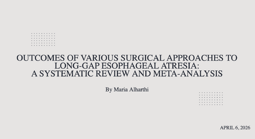 OUTCOMES OF VARIOUS SURGICAL APPROACHES TO LONG-GAP ESOPHAGEAL ATRESIA: A SYSTEMATIC REVIEW AND META-ANALYSIS LECTURE
 -
quote from "Pediatric Colorectal Conundrums: Case Studies: From Fundamental to Advanced (Pediatric Colorectal Surgery)" by Marc Levitt, Thomas Xu, Hussein Wissanji - "A recent study from the ARM-Net registry highlights the need for sufficient screening and vigilant management of high-grade VUR in ARM patients. This study underscores the fact that despite normal kidneys on US, a significant proportion of patients may still have high-grade VUR, necessitating prophylaxis. All patients with ARM should have a newborn screen renal US and, in most, a follow-up VCUG should be done to assess for VUR."
-
until
On Monday 13/4, European Society for Paediatric Gastroenterology, Hepatology and Nutrition (ESPGHAN) Webinar - Paediatric Liver Disease: Genetic and Nutrition. This webinar is organized by the ESPGHAN Special Interest Groups on Childhood Obesity and Fatty Liver. Starting at 2 pmGMT , 4 pm Cairo time Register through the link: https://zoom.us/webinar/register/WN_bzvAwkMLQAGgIHWXThLF6Q#/registration -
until
On Monday 13/4, Surgical Management of Acute and Chronic Pancreatitis in Children with Dr. Juan Gurria. This webinar is provided by Cincinnati Children’s Hospital. Starting at 3 pm GMT, 5 pm Cairo time Register through the link: https://globalcastmd.com/wp-admin/admin-ajax.php?action=frm_forms_preview&form=overview-of-the-surgical-management-of-acute-and-chronic-pancreatitis-in-children&theme=1 -
until
On Tuesday 14/4, The Pan African Pediatric Surgical Association (PAPSA) will be holding their first webinar of the PAPSA monthly webinar series in collaboration with CLIRNET. This month talk will be deliveredby Prof. Ashraf Hafez on Bladder extrophy Management. Starting at 3 pm GMT, 5 pm Cairo time Register through the link: https://doctor.clirnet.com/share/session/23991/glo_papsa_bladder/ -
until
On Tuesday 14/4, Cure4u collaborative with the European Reference Network (ERN) are conducting their monthly colorectal cases discussions. This month features a special joint session with the German PediatricSurgery Association. Starting at 4 pm GMT, 6 pm Cairo time Registerthrough the link: https://www.cureforu.com/subscriptions/new -
until
On Wednesday 15/4, European Society for Paediatric Gastroenterology, Hepatology and Nutrition (ESPGHAN) Webinar - Pediatric Polyposis Syndromes: From Genetics to Endoscopic Management. Presented by: Tomas Attard, UK. This webinar is part of the 2026 Young ESPGHAN Webinar Series. Starting at 2 pm GMT , 4 pm Cairo time Register through the link: https://zoom.us/webinar/register/WN__lBcE5HLRsi1_n-kFxEREQ#/registration -
until
On Tuesday 21/4, Joint CGA-IGC & NASPGHAN webinar on “The incidental adenoma in the young and adolescent population”. Presented by Aparajita Singh and Brett Hoskins. Starting at 4 pm GMT , 6 pm Cairo time Register through the link; https://www.addevent.com/event/rghvmbwyxcwl?fbclid=IwY2xjawRAI4xleHRuA2FlbQIxMQBzcnRjBmFwcF9pZA80MDk5NjI2MjMwODU2MDkAAR7y6H_K0ZkRrlBRlvXqKTn2yDBPWOBD6oCVSUVDfaRtJC33KSTKJl1F1AZupw_aem_yTFiY5mRqx-FtoZD2FLGtQ -
until
On Monday 27/4, Case-based discussion webinar on the management of neuroblastoma. Presented by Drs. Katherine Somers, Cara Morin, Juan Gurria, and Meera Kotagal. Starting at 2,30 pm GMT, 4,30 pm Cairo time Register through the link: https://globalcastmd.com/wp-admin/admin-ajax.php?action=frm_forms_preview&form=clinical-and-research-update-neuroblastoma&theme=1 -
quote from "Pediatric Colorectal Conundrums: Case Studies: From Fundamental to Advanced (Pediatric Colorectal Surgery)" by Marc Levitt, Thomas Xu, Hussein Wissanji - "If the frozen section reveals active enterocolitis in a ganglionated bowel section without nerve hypertrophy, diversion should be considered as active enterocolitis can increase the risk of anastomotic complications such as stricture and dehiscence."
-
quote from "Pediatric Colorectal Conundrums: Case Studies: From Fundamental to Advanced (Pediatric Colorectal Surgery)" by Marc Levitt, Thomas Xu, Hussein Wissanji - "During the operation for HD, frozen section biopsies are used. Frozen section biopsies cannot diagnose HD, but they can rule it out. They only capture one section of the bowel and may be useful intraoperatively for confirming the presence of ganglionated bowel for an anastomosis or a stoma site. Permanent sections are the gold standard for diagnosis and are used to determine transition zones. As previously mentioned, the distribution of ganglion cells is irregular at the transition zone, and multiple slices (>100) need to be evaluated circumferentially as a single slice could capture one of the few ganglion cells within a transition zone and falsely lead to a diagnosis of normal colon or the reverse could occur. Frozen section might not see ganglion cells which in fact are nearby, and have the clinician draw the wrong conclusion that the bowel is aganglionic. Frozen section is subject to artifact from cracking of the tissue and can lead to the pathologist not being able to see ganglion cells which are in fact present. Permanent sections also offer a better appreciation and measurement of nerve hypertrophy. They take a longer time to process and are not available intraoperatively"
-
quote from "Pediatric Colorectal Conundrums: Case Studies: From Fundamental to Advanced (Pediatric Colorectal Surgery)" by Marc Levitt, Thomas Xu, Hussein Wissanji - "Anorectal manometry is sometimes used to diagnose HD; however, its diagnostic accuracy is limited in children under the age of 12 months. It is a helpful adjunct in diagnosing older children who struggle with constipation due to a delayed diagnosis of HD. One of the characteristics of HD is that patients cannot relax their internal anal sphincter in response to rectal distension, otherwise known as the rectoanal inhibitory reflex (RAIR). Anorectal manometry can screen for the absence of this reflex, which is typical of HD. If the RAIR is absent, a rectal biopsy to assess for HD should be done."
-
Quote from "Pediatric Colorectal Conundrums: Case Studies: From Fundamental to Advanced (Pediatric Colorectal Surgery)" by Marc Levitt, Thomas Xu, Hussein Wissanji - "The main differential of surgical diagnoses for an infant with a distal bowel obstruction are: • HD • Neonatal small left colon • Anorectal malformation • Jejuno-ileal atresia • Meconium ileus • Meconium plug syndrome Some medical causes of colonic distension include: • Hypothyroidism • Magnesium sulfate effect • Opiate effect • Milk protein allergy"
-
-
-
-
-
quote from "Pediatric Colorectal Conundrums: Case Studies: From Fundamental to Advanced (Pediatric Colorectal Surgery)" by Marc Levitt, Thomas Xu, Hussein Wissanji - "Vaginal loss or acquired vaginal atresia, like urethral loss, occurs from ischemia related to excessive dissection and tension. When UG separation is required, it is important to maintain the vaginal blood supply from the lateral attachments bilaterally. For this reason, older techniques such as the vaginal switch have been abandoned. Optimal mobilization of the vagina is achieved by full separation from the common channel and fully dividing the central pelvic and retroperitoneal attachments posteriorly. The blood supply then depends on the round ligaments from both sides. A vaginal length of 4 cm is predictive of a successful native vaginal pull-through. When the Müllerian structures are smaller or absent, delayed vaginal reconstruction or bowel vagina replacement should be considered. If a bowel neovagina is used to bridge the gap to the perineum, this could be removed later in life with a pull-through, at that time, of the native vagina."
-
quote from "Pediatric Colorectal Conundrums: Case Studies: From Fundamental to Advanced (Pediatric Colorectal Surgery)" by Marc Levitt, Thomas Xu, Hussein Wissanji - "Urethral loss or urethrovaginal fistula can occur due to ischemia related to over-dissection of the common channel. If the surgeon underestimates the common channel length and begins a TUM dissecting the anterior urethra but then realizes intraoperatively that there is insufficient mobilization to reach the perineum, changing to a UG separation and dissecting the posterior urethral wall risks significant ischemia of the now circumferentially dissected common channel. Such complications can lead to an acquired bladder neck closure and the need for a Mitrofanoff."
-
quote from "Pediatric Colorectal Conundrums: Case Studies: From Fundamental to Advanced (Pediatric Colorectal Surgery)" by Marc Levitt, Thomas Xu, Hussein Wissanji - "When the common channel is <3 cm, it is possible to mobilize the UG complex as a unit to the perineum, the TUM. In general, a urethral length of >1.5 cm is required to avoid urine leakage and to avoid pulling down the bladder neck out of the urogenital diaphragm with the mobilization. When the length of the common channel is >3 cm or the urethra is <1.5 cm, a UG separation is utilized, leaving the common channel untouched and adding to it the native urethra. With the UG separation, the vagina and rectum are dissected off of the common channel and mobilized to bring them each down to the perineum independently. When the common channel is >3 cm, a UG separation is required to gain the necessary mobilization for the vagina to reach the perineum. After separation, the common channel is repaired, and the urethra and common channel together become the new urethra now with adequate length."
-
-
quote from "Pediatric Colorectal Conundrums: Case Studies: From Fundamental to Advanced (Pediatric Colorectal Surgery)" by Marc Levitt, Thomas Xu, Hussein Wissanji - "In patients who have had a vaginal replacement, the most common long-term complication is stenosis of the neovagina. This can often be diagnosed with an exam under anesthesia and managed with dilations but may require a revision. If bowel interposition was used for vaginal replacement, patients may experience prolapse of the interposition, excessive mucus production, bleeding, mal-odor, and dyspareunia. Malignancy of the bowel graft has been reported, as has inflammatory bowel disease, in the segment. These are the main reasons why bowel neovaginas nowadays are avoided. These complications are eliminated in patients bridged with buccal mucosa."
-
Quote from "Pediatric Colorectal Conundrums: Case Studies: From Fundamental to Advanced (Pediatric Colorectal Surgery)" by Marc Levitt, Thomas Xu, Hussein Wissanji - "For patients with uterine abnormalities, obstruction of menses is possible, and surgical correction may become necessary. Pregnancies in patients with vaginal anomalies are often considered high risk, and delivery options need to be discussed with maternal fetal medicine. The pelvic floor muscles are often not normally developed or have been iatrogenically altered, making vaginal delivery after pregnancy more traumatic. This is especially true if the perineal body was divided during reconstruction. Most patients with a history of anorectal and/or vaginal malformations are advised to use c-section as the mode of delivery."
-
quote from "Pediatric Colorectal Conundrums: Case Studies: From Fundamental to Advanced (Pediatric Colorectal Surgery)" by Marc Levitt, Thomas Xu, Hussein Wissanji - "Low ARMs, like recto-vestibular and perineal fistula, tend to be associated with congenital rectal dilation above the fistula. The dilated rectum is prone to constipation, and early bowel management is recommended. Bowel management in this population utilizes laxatives and fiber to keep the rectum empty and avoid distention of the dilated rectum. The pelvic floor and sphincter muscle may be more robust in these distal malformations; however, incontinence is still possible, especially in the setting of an associated spinal or sacral anomaly. Pelvic floor physiotherapy can assist with developing the ability to fully evacuate stool and hold onto the stool, both needed to improve continence. Rectal or ante-grade enemas can be helpful in achieving social continence when laxatives are ineffective and can be used as a bridge until the child achieves the capacity for voluntary bowel movements."
-
Cardiac tamponade after subclavian vein porta cath insertion, case for discussion sent by a colleague male patient 2 yrs old presented with ACUTE LYMPHOBLASTIC LEUKEMIA, presented for porta cath insertion, with low platelets count. After correction of thrombocytopenia, under GA, percutaneous needle inserted to cannulate the right subclavian vein, infra clavicular, succeeded from first trial without issues, then guide wire inserted and position checked with fluoroscopy in the right atrium, followed by catheter insertion smoothly, catheter connected to the porta cath chamber with good inflow and outflow no resistance. Suddenly during skin closure patient became bradycardic then arrested, CPR started and patient returned after few cycles. We repeated portable chest xray in OR while patient still intubated and no clear evidence of hemo or pnemo thorax. Yet patient still hypotensive and about to arrest, so needle decompression tried to right chest nothing came out followed by right chest tube and no blood or air came out. Bed side echo done and cardiac tamponade found. cardiac surgery colleague contacted to join then we inserted subxyphoid needle and aspirated blood 150 cc. Decision made with cardiac surgery to proceed with median sternotomy, pericardium opened blood came out marked amount, cardiac massaging started, heart started beating, on assessment of heart no cardiac injury found and normal svc and ivc, Yet injury found at the innominate veins bifarcation which is repaired, and sternotomy closed. Patient survived but with neurological sequel. the injury could be due to the guide wire or due to the catheter, the question, is there any thing could be done to avoid this rare complication of central venous insertion?, and weather there is malpractice in the management of this patient ?



.jpeg.f6d56979672ea500da73a7fad634ec2d.jpeg)
.jpeg.313dab3bf5ee0968feede8b3093fab95.jpeg)
.jpeg.8d10d3102de72044bd5e3413a6f0cde2.jpeg)
.jpeg.797090e863a21938599f57984a37d775.jpeg)
.jpeg.e0094ce31797c2af82eb5e6595b2d3d3.jpeg)