


admin
Administrators
-
Joined
-
Last visited
Everything posted by admin
-
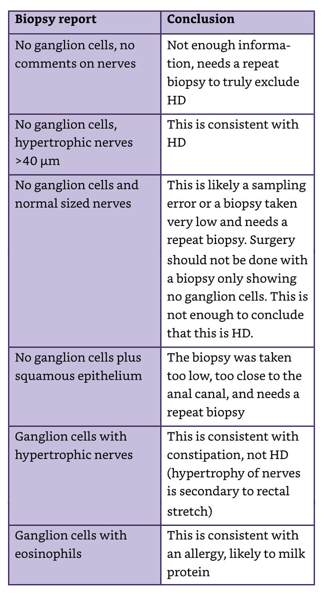
quote from "Pediatric Colorectal Conundrums: Case Studies: From Fundamental to Advanced (Pediatric Colorectal Surgery)" by Marc Levitt, Thomas Xu, Hussein Wissanji - "A colonic resection based solely on a frozen section must be avoided in patients who have an aganglionic zone proximal to the left colon. This is because proximal to the splenic flexure you cannot expect to see hypertrophic nerves (the sacral plexus does not innervate that bowel). Therefore, you are completely dependent upon your pathologist identifying ganglion cells on frozen section which is prone to sampling bias /false negatives. This is due to artifact in the tissue that can occur from the freezing process. Consequently, you risk resection of good bowel that was incorrectly deemed aganglionic by frozen section."
-
-
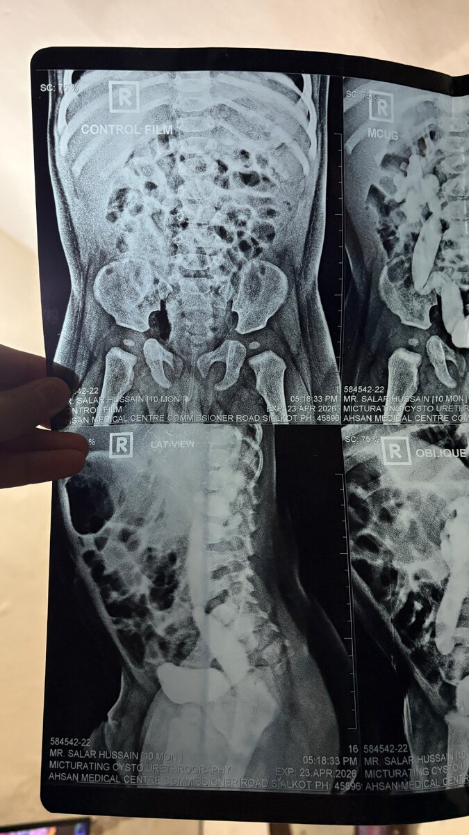
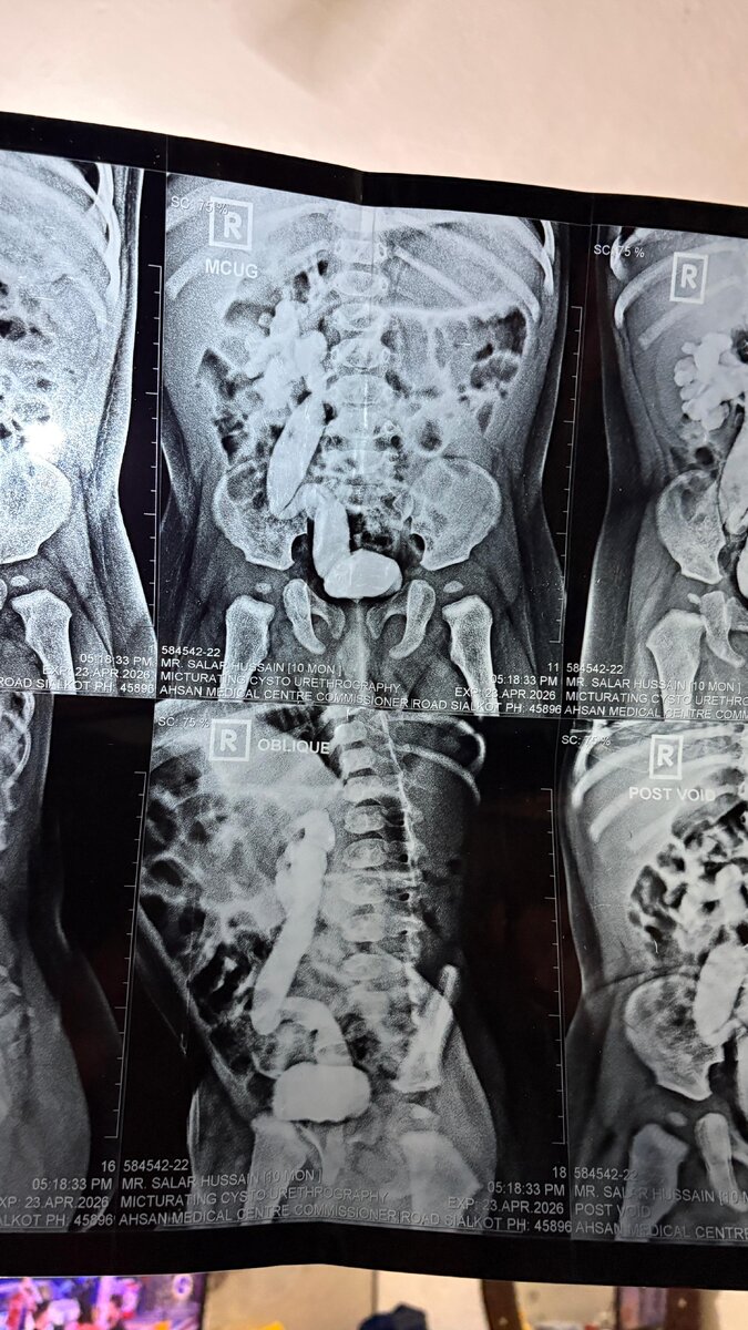
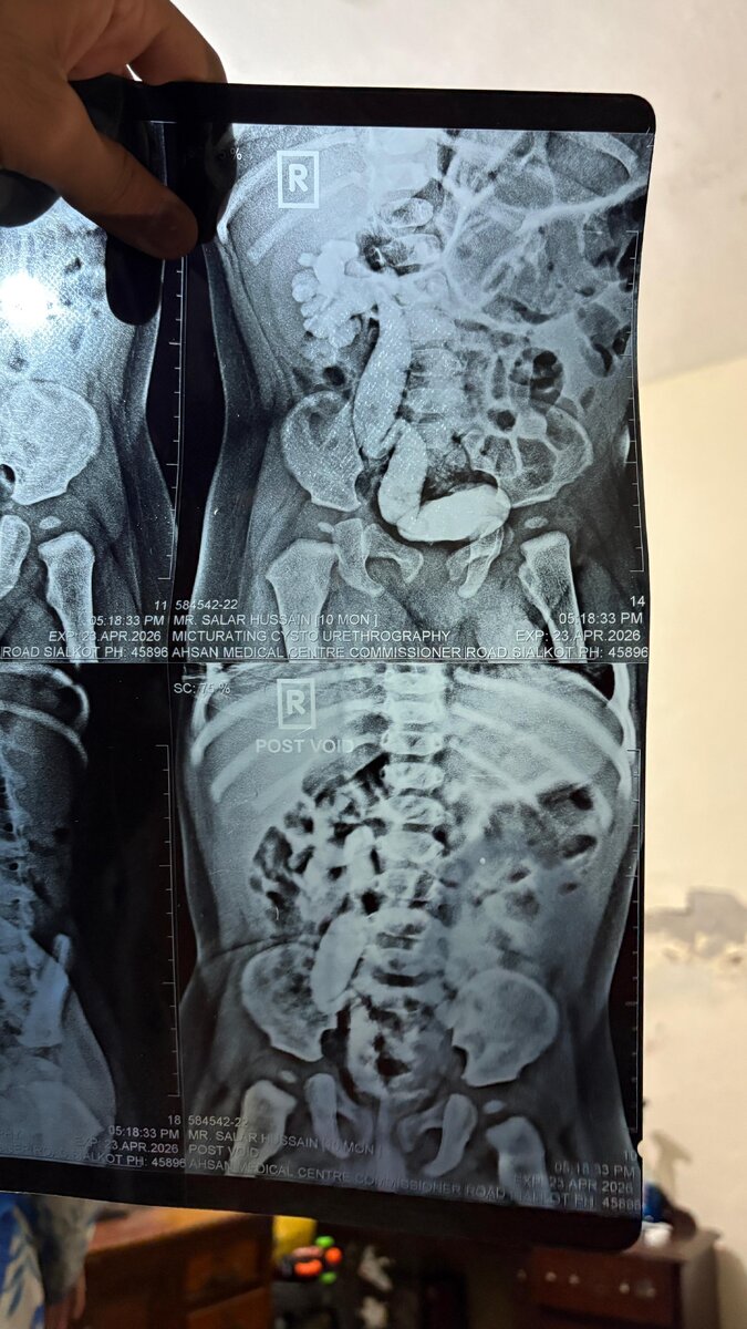
case from Dr Usman Akram What will be the best plan according to you in this case ?
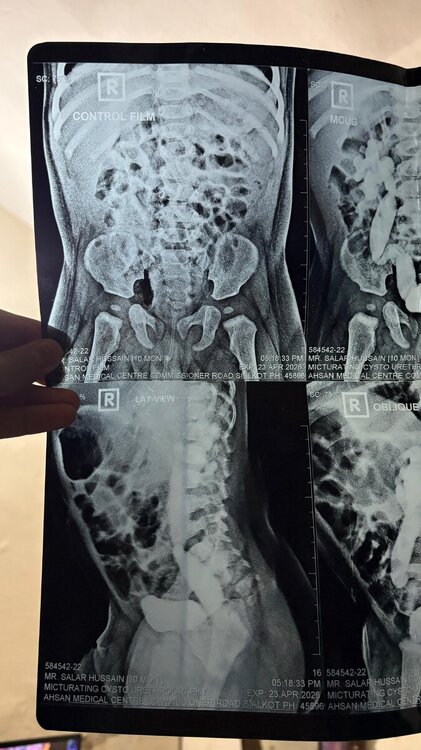
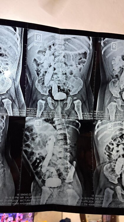
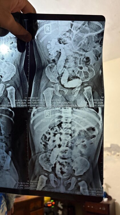
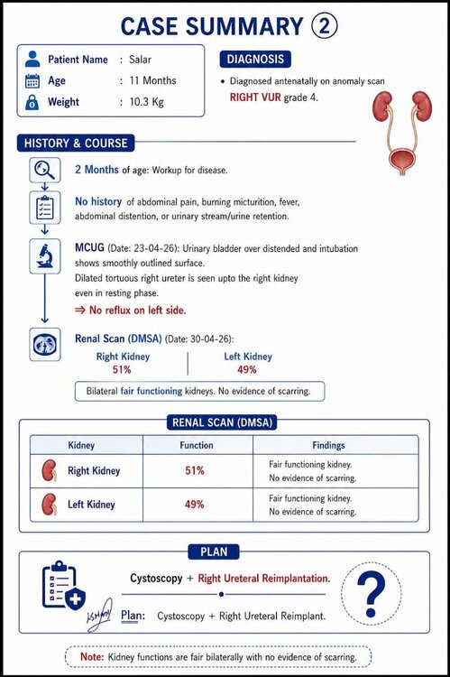 quote from "Pediatric Colorectal Conundrums: Case Studies: From Fundamental to Advanced (Pediatric Colorectal Surgery)" by Marc Levitt, Thomas Xu, Hussein Wissanji - A technically correct high-pressure distal colostogram should provide data on the following six key components: 1. The mucous fistula site. 2. The amount of bowel length available for the pull-through (AP view). 3. The rectal end and communication with the urinary tract (if present) (lateral view). 4. The tip of the sacrum. 5. The urethra and bladder (if possible). 6. An anal marker.quote from "Pediatric Colorectal Conundrums: Case Studies: From Fundamental to Advanced (Pediatric Colorectal Surgery)" by Marc Levitt, Thomas Xu, Hussein Wissanji - 1. For all anal stenosis (and rectal atresia) cases you must check for a presacral mass. 2. For any such presacral mass, always make sure there is no dural component (pelvic plus spinal MRI) and, if present, involve neurosurgery. 3. If Currarino syndrome, genetic testing is warranted for the patient and first-degree relatives. 4. For management, removal of the mass is key which will likely solve much of the constipation. 5. If the anus is narrow that must be treated; an anal canal sparing technique can accomplish this. Occasionally, dilation alone can work. 6. With mass removal the colon should improve, although in such a case antegrade flush access with a Malone may be beneficial to promote daily colonic emptying. 7. Whether the colon will recover is a key question, but it can be decided later whether a colon resection is required.
until
On Monday 11/5, North Thames Paediatric Network (NTPN ODN)– Pediatric Robotic-Assisted Surgery (RAS) clinical educational forum and training webinar for paediatric clinicians across the North Thames area, focusing on surgical practice for nursing, medical, AHP and HCSW staff involved in paediatric surgery. Starting at 9 am GMT , 12 pm Cairo time Register through the link https://www.eventbrite.co.uk/e/ntpn-odn-paediatric-robotic-assisted-surgery-ras-morning-tickets-1984388060106?utm-campaign=social&utm-content=attendeeshare&utm-medium=discovery&utm-term=listing&utm-source=cp&aff=ebdsshcopyurluntil
On Tuesday 12/5, The Pan African Pediatric Surgical Association (PAPSA) will be holding their monthly webinar series in collaboration with CLIRNET. This month talk will be delivered by Prof. Sameh Shehata on Management of Intra-abdominal Testes Registration link https://doctor.clirnet.com/share/session/23990/GLO_Papsa_Management/until
On Tuesday 12/5, Cure4u collaborative with the European Reference Network (ERN) are conducting their monthly colorectal cases discussions. Starting at 4 pm GMT, 7 pm Cairo time Register through the link: https://www.cureforu.com/subscriptions/newuntil
On Wednesday 20/5, International pediatric live surgery online group holding the bimonthly course. This course topic is on “Pancreatic Surgery”. Starting at 7,30 am GMT , 10,30 am Cairo time Register through the link; https://www.pediatriclivesurgery.it/until
On Wednesday 27/5, Surgery - Radiology - Pathology Rounds with The Montreal Children's Hospital sponsored by The Henderen Project. Starting at 12 pm GMT , 3 pm Cairo time Register through the link: https://us02web.zoom.us/webinar/register/WN_NbRmPi8sRnq0dJj-wF192g#/registrationuntil
On Thursday 28/5, The monthly international colorectal web meeting by Colorado children’s hospital sponsored by The Henderen Project Starting at 4 pm GMT , 7 pm Cairo time Register through the link; https://www.hendrenproject.org/case/about-international-colorectal-web-meetingsRegarding PSARP for recto-bulbar fistula quote from "Pediatric Colorectal Conundrums: Case Studies: From Fundamental to Advanced (Pediatric Colorectal Surgery)" by Marc Levitt, Thomas Xu, Hussein Wissanji - "The key maneuver required is to open the posterior rectal wall and carefully inspect the anterior wall for any connections and, if a fistula is found, then carefully mobilize the anterior rectum off the urethra."quote from "Pediatric Colorectal Conundrums: Case Studies: From Fundamental to Advanced (Pediatric Colorectal Surgery)" by Marc Levitt, Thomas Xu, Hussein Wissanji - "while the AP view might misleadingly suggest an underdeveloped sacrum, the lateral view typically provides a more accurate representation. The pelvic tilt can distort the appearance of the sacrum and coccyx in the AP view, leading to a falsely low sacral ratio. The lateral view, however, allows for a more reliable calculation by minimizing distortions caused by patient positioning"quote from "Pediatric Colorectal Conundrums: Case Studies: From Fundamental to Advanced (Pediatric Colorectal Surgery)" by Marc Levitt, Thomas Xu, Hussein Wissanji - "It is crucial not to chase the anteriorly directed fistula toward the scrotum, as it runs parallel to the urethra. Attempting to trace this fistula surgically could lead to significant complications, including spongiosum bleeding and urethral injury. Instead, the focus should be on mobilizing the distal rectum and avoiding dissection of the anterior rectal wall to avoid a urethral injury."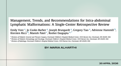 intra-abdominal lymphatic malformation management lecture


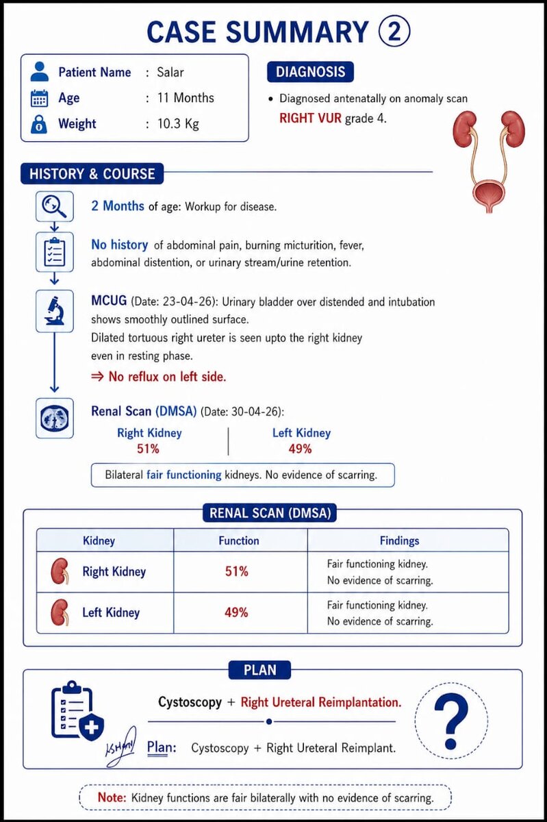

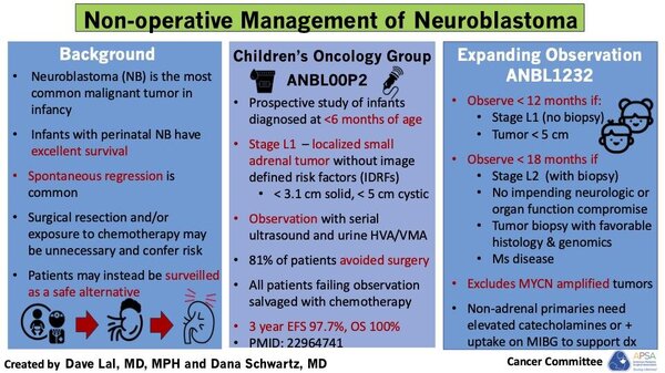
.jpeg.e02b45d0cb1223bfe689b01c89dd2569.jpeg)
.jpeg.676ad9f93d6992056251290480a8a2cf.jpeg)
.png.956f202b98526574470746c451b204a6.png)
.jpeg.8dd69ad1a3f90205bf31963552948f5b.jpeg)
.jpeg.67c8b22c64eaeb2da5793e1b34b00502.jpeg)
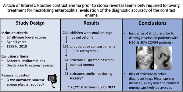
Account
Navigation
Search
Configure browser push notifications
Chrome (Android)
- Tap the lock icon next to the address bar.
- Tap Permissions → Notifications.
- Adjust your preference.
Chrome (Desktop)
- Click the padlock icon in the address bar.
- Select Site settings.
- Find Notifications and adjust your preference.
Safari (iOS 16.4+)
- Ensure the site is installed via Add to Home Screen.
- Open Settings App → Notifications.
- Find your app name and adjust your preference.
Safari (macOS)
- Go to Safari → Preferences.
- Click the Websites tab.
- Select Notifications in the sidebar.
- Find this website and adjust your preference.
Edge (Android)
- Tap the lock icon next to the address bar.
- Tap Permissions.
- Find Notifications and adjust your preference.
Edge (Desktop)
- Click the padlock icon in the address bar.
- Click Permissions for this site.
- Find Notifications and adjust your preference.
Firefox (Android)
- Go to Settings → Site permissions.
- Tap Notifications.
- Find this site in the list and adjust your preference.
Firefox (Desktop)
- Open Firefox Settings.
- Search for Notifications.
- Find this site in the list and adjust your preference.